

MICROSCOPES AND SYSTEMS FOR IMAGE ANALYSIS / Inverted Microscopes

RESEARCH microscopes
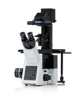
OLYMPUS IX73 MICROSCOPE
Olympus IX73 is a semi-motorized inverted microscope designed to meet a variety of research needs. With two microscope frame options and optional modules to expand functionality, the IX73 is ideal for a changing research environment.
Read More
IX73: Two-deck System. The IX73 modular two-tier system can be combined with coded or motorized units for maximum functionality expansion.
IX73: One-deck System. A microscope designed with efficiency in mind. Suitable for documentation, routine research and other tasks.
Thanks to the high resolution, users have the opportunity to observe clear and bright images of excellent quality.
Olympus UIS2 infinity-corrected optics provide high optical throughput with a wide range of lenses. UIS2 optics are characterized by wide chromatic correction and allow obtaining primary images with high resolution and high signal/noise ratio, regardless of the observation method. The wide viewing angle and the Fly-Eye lens system provide uniform fluorescent images and allow the use of SCMOS cameras with large sensors.
Apochromatic objectives allow for high-resolution phase-contrast and fluorescence observations.
Apochromatic phase contrast lenses (UPLSAPO100XOPH, PLAPON60XOPH) allow obtaining high-precision images without image shift even when observing phase contrast and fluorescence simultaneously. This eliminates the need to change lenses when changing observation methods.
The IXplore Standard system was developed on the basis of this microscope. Designed for simple multi-color fluorescence imaging and routine experiments, it is easy to operate and produces excellent publication quality images. A number of coded module options facilitate accurate and reproducible results.
The manual stage is equipped with a smooth positioning system that allows easy tracking of cells even at high magnification.
The observation limit settings fix the stage and help maintain its position, including during the application of reagents, even if the stage is accidentally touched.
Using the table fixing plate and screws, you can remove the 35 mm dish from the table, transfer it to the incubator and return it to the exact previous position.
A wide range of additional devices are available for modernization, including: 8-position motorized or coded turret with fluorescent mirrors; 6-position motorized or coded turret lens head; motorized universal condenser with long working distance.
Lenses of the LUCPLFLN series are well suited for observations using plastic plates. Lenses allow for high-resolution observation of the process of cell proliferation and provide improved contrast over a large area. This gives you the ability to get images through plastic-bottomed dishes in addition to glass.
Fluorescent mirror units are equipped with filters with a specially designed coating that absorbs more than 99% of scattered light. This reduction in reflection and the high transmittance of the mirror blocks help produce fluorescence images with a high signal-to-noise ratio.
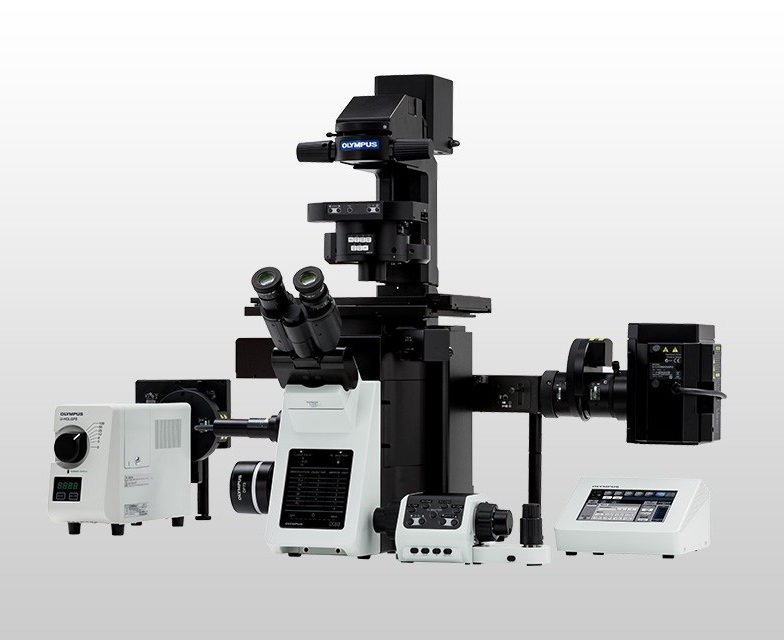
OLYMPUS IX83 MICROSCOPE
Olympus IX83 is a fully motorized system designed for various research tasks. With add-on modules providing enhanced functionality, both microscope variants enable a variety of imaging techniques, from casual documentation to long time-lapse imaging and other complex techniques.
IX83: Two-deck System. Provides high-speed, fully automated device selection during real-time examinations and advanced imaging. The two-tier configuration provides excellent expandability.
IX83: One-deck System. Intelligent motorized microscope compatible with TruFocus, creating a new standard for live cell imaging.
Read More
The unique open frame of the IX83 microscope provides free access to the light path, making it easy to add or replace modules. Different deck modules can be easily swapped out to add or remove features as needed. With a simple plug-in design, the TruFocus Z drift compensator module can easily be added to any IX83 system to maintain continuous focus during a long interval shooting experiment.
Olympus UIS2 infinity-corrected optics provide high optical throughput with a wide range of lenses. UIS2 optics are characterized by wide chromatic correction and allow obtaining primary images with high resolution and high signal/noise ratio, regardless of the observation method.
The IXplore Pro system was developed on the basis of this microscope. for automated multidimensional observation and simple experiment setup, simplifying data acquisition and seamless acquisition of panoramic multichannel images.
The graphical experiment manager (GEM) of cellSens Dimension software offers fully automated multidimensional observation (X, Y, Z, T, wavelength and position) and simplifies experiment setup. Macro functions such as performing deconvolution processing can be defined to improve GEM performance.
A multi-point focus map enables automatic focusing of wide areas of the image with multiple lenses, making focusing easier when moving around samples.
The fluorescent illuminator includes a “Fly-Eye” lens system for even distribution of light. The system provides bright and uniform illumination of the entire field of view, which is well suited for stitching images.
The software also includes real-time 2D deblurring for preview and image acquisition, allowing for better focusing of thick specimens. To further improve detail, TruSight deconvolution is available to remap out-of-focus light. TruSight uses a constrained iterative deconvolution algorithm to improve resolution, contrast and dynamic range at industry-leading speed through GPU processing. To improve the efficiency of the experiment, the deconvolution processing can be defined as a GEM macrofunction.
Images can be easily converted to statistically significant data using CellSens software. The software includes region of interest, phase analysis and cell counting capabilities. Export measurement data to Microsoft® Excel® or a CellSens workbook with one click.
LABORATORY microscopes

OLYMPUS CKX53 MICROSCOPE
With improved image quality and ease of use, the Olympus CKX53 microscope provides consistent performance and efficiency in a variety of research applications, including live cell observation, cell sampling and processing, image capture, and fluorescence observation.
The microscope’s long-life LED system and iPC (integrated phase contrast) system provide the ability to obtain clear and high-contrast images with a wide field of view. In addition, the inversion contrast (IVC) technique provides a clear three-dimensional image.
The CKX53 microscope is equipped with a standardized camera port, so it can be connected to an Olympus camera, allowing users to quickly obtain clear images in bright light, phase contrast, inversion contrast and fluorescence imaging modes.
Read More
When observing fluorescence, a wide range of fluorescent dyes can be used by changing the microscope mirror. Due to the increased filtering ability of the mirror unit, high-contrast fluorescence images with a high signal-to-noise ratio can be obtained, even when the fluorescence is relatively weak. In addition, the microscope’s LED and LDP light source provide clear, bright fluorescence observation.
The microscope’s width and easily removable condenser allow viewing of containers such as multilayered tissue flasks up to 190 mm (7.5 inches) high.
The depth of field of the PLCN4X objective makes it easy to observe the cells in the bottom two layers of the multilayer flask.
The high contrast of the CKX53 iPC system provides sharp images without the need to change the annular gap from a 4x to a 40x objective. This simplifies cell observation for a faster and more convenient workflow.