

MICROSCOPES AND SYSTEMS FOR IMAGE ANALYSIS / Confocal microscopes

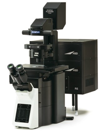

MICROSCOPE OLYMPUS FLUOVIEW FV3000
The FLUOVIEW FV3000 series of confocal laser scanning microscopes meets some of the most difficult challenges in modern science. Featuring the high sensitivity and speed required for live cell imaging as well as deep tissue observation, the FV3000 confocal microscope enables a wide range of imaging modalities, including macro-to-micro imaging, super resolution microscopy, and quantitative data analysis.
Using proprietary spectral detection technology, the FV3000 confocal microscope’s TruSpectral detectors combine high sensitivity with spectral flexibility to detect even the dimmest fluorophores.
- TruSpectral technology achieves up to 3X more light transmission vs. traditional spectral detection technology by implementing the volume phase holographic (VPH) diffraction grating.
- Offers independently adjustable channels to optimize signal detection for each individual fluorophore. Variable barrier filter mode provides simultaneous four-channel image acquisition with up to sixteen channels in sequential mode.
- Lambda scanning mode enables accurate spectral unmixing of complex overlapping fluorescent signals.
Our new FV3000 Red near-infrared (NIR) solution further extends the FV3000 microscope’s wavelength detection capabilities to the NIR region of up to 890 nm through a suite of carefully engineered NIR upgrades:
- 730 nm or 785 nm lasers and an NIR-sensitive 1- or 2-channel GaAs detectors enable up to 6 channels for multiplexed imaging from violet to NIR (400 nm–890 nm).
- TruFocus Red enables stable and gentle NIR time-lapse imaging.
- X Line objectives offer the widest range of chromatic correction, from 400 nm – 1000 nm, for exceptional color accuracy during multicolor fluorescence imaging.
The FV3000 Hybrid Scanner provides two scanners in one for enhanced confocal imaging capabilities.
- The FV3000RS hybrid scan unit uses a galvanometer scanner for precision scanning as well as a resonant scanner, ideal for high-speed imaging of live physiological events
- Capture video-rate images with a large FOV using the resonant scanner, featuring speeds from 30 frames per second (fps) at FN 18 all the way up to 438 fps using clip scanning
- Use the resonant scanner to observe fast phenomena, such as a beating heart, blood flow, or calcium ion (Ca2+) dynamics inside cells
- Switch between the galvanometer scanner and resonant scanner with the click of a button