

MICROSCOPES AND SYSTEMS FOR IMAGE ANALYSIS / Upright Microscopes

Laboratory microscopes
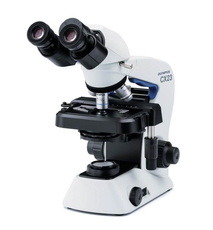
MICROSCOPE OLYMPUS CX23
The Olympus CX23 is a microscope designed to solve everyday (routine) tasks in clinical diagnostic practice. This is a professional optical microscope with a 360° rotatable binocular objective, with an eyepiece tilt angle of 30°.
The device is equipped with a modern optical system UIS2 (Universal Infinity System type 2), which significantly improves the flatness of the image, creating high quality and simultaneous sharpness throughout the image field. Infinity-corrected plan-achromatic lenses of 4x, 10x, 40x and 100xO provide crisp, high-contrast images across the entire field of view. The built-in powerful LED light source with a long service life and low power consumption provides bright illumination when working with the sample.
The СХ23 microscope can be equipped with a trinocular attachment (360° rotatable, 30° eyepiece tilt angle) to enable easy image capture and recording.
Designed to improve workflow, the CX23 direct microscope requires minimal setup.
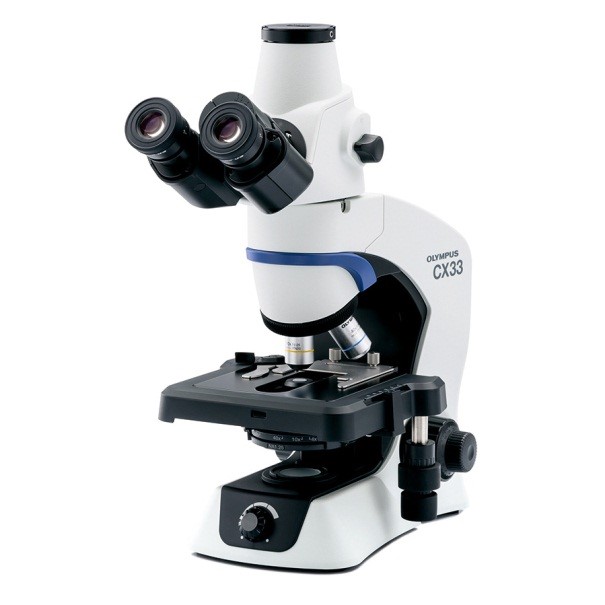
MICROSCOPE OLYMPUS CX33
The Olympus CX33 microscope is a great option when you need to use only light and dark fields. The low, inwardly rotating, four-position turret head, stage, focusing lock, and sample holder make the CX33 well suited for everyday observations in one simple configuration. The microscope has a camera port for digital imaging and is equipped with specially designed plan-achromatic objectives that provide clear, high-fidelity images over a wide field of view.
The CX33’s LED lighting provides several immediate and long-term benefits:
- Creates daylight conditions so that samples can be viewed in their natural colors.
- Constant color temperature at any brightness, so you don’t have to waste time adjusting when changing brightness
- With a long service life (60,000 hours), the brightness level remains stable throughout.
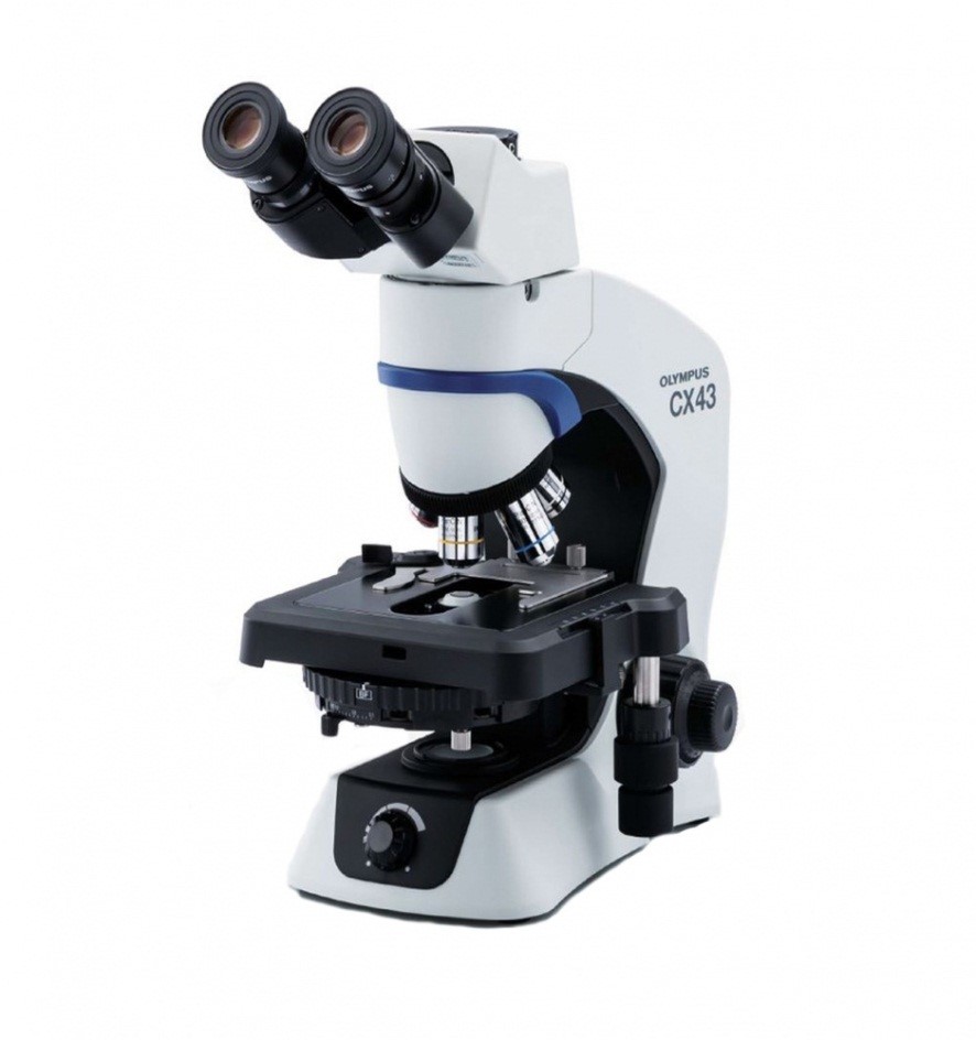
MICROSCOPE OLYMPUS CX43
The Olympus CX43 microscope allows users to feel comfortable while working. The microscope’s frame conforms comfortably to the hands, and the positioning of the controls increases ergonomics for improved work efficiency.
The color temperature of the microscope’s LED illumination creates daylight conditions, so samples can be viewed in their natural colors. The LEDs have a long lifespan of 60,000 hours, which helps to reduce costs, and the brightness level remains stable throughout the life of the LED.
The microscope uses plan-achromatic objective lenses that are compatible with all UIS2 objectives, providing crisp, high-fidelity images over a wide field of view.
The Olympus CX43 microscope can be equipped with a fluorescence function. Observing fluorescence is easy. Simply insert the compact fluorescence illuminator into the microscope body and set the condenser turret to the FL position to reduce background noise caused by stray light coming from the top of the condenser.
The Olympus CX43 also has a camera port for digital imaging.
Research microscopes

MICROSCOPE OLYMPUS BX43
The Olympus BX43 microscope is robust and easy to use. The BX43 allows you to choose between standard and advanced configurations depending on your needs. Modular components, such as ergonomic viewing tubes and slides, make it easy to adapt the microscope to your application.
The BX43 microscope uses a high-quality LED with a luminous intensity equivalent to a 30 W halogen lamp. The long-life LED ensures a constant color temperature at any brightness level.
Thanks to the optional low-magnification condenser, users can change the objective magnification from 2X to 100X (dry) without changing the condenser or moving the upper lens.
The microscope can be mounted with 4 to 7 objective lenses with magnifications from 2X to 100X UIS2 standard.
Eight fluorescence mirror blocks can be attached to the microscope to enable multi-color fluorescence observations. High performance filters ensure efficient and bright fluorescence images.
High-contrast phase imaging enables direct observation of the internal environment of cells and live bacteria. To observe phase contrasts from 10X to 100X, the UPLFLN-PH or PLN-PH objective lens must be used. With the U-PCD2 Phase/Dark Field Condenser, users can view samples in either bright field or dark field. Simultaneous observation with reflected light fluorescence microscopy is possible.
Also available are a condenser for polarized light, a turret head, a rotary slide, objective lenses, and an analyzer for detecting uric acid and crystals.
The light intensity control adjusts the brightness to the specific objective. Homogeneous brightness is achieved from low to high magnification levels, eliminating the need to adjust the light intensity, which reduces eye fatigue.
The microscope can be fitted with a trinocular tube to equip it with a digital camera for easy image capture and recording.

MICROSCOPE OLYMPUS BX46
The Olympus BX46 microscope is an improved version of the Olympus BX43 microscope, with improved ergonomics and enhanced functionality. It provides a comfortable environment for users when performing standard pathological and cytological examinations. The BX46 microscope has a low frame position and a turret with inwardly oriented objectives, which, combined with a fixed low-position stage, allows for easy and quick microsection changes.
The color rendering index of the LED is similar to a halogen lamp with a daylight filter, but unlike a halogen light source, the lifetime of the LED is estimated at 20,000 hours.
Thanks to the optional low-magnification condenser, users can change the objective magnification from 2X to 100X (dry) without changing the condenser or moving the upper lens.
The microscope is equipped with a 5-lens revolver that can accommodate UIS2 objective lenses from 2X to 100X.
Eight fluorescence mirror blocks can be attached to the microscope to enable multi-color fluorescence observations, which is achieved through high-performance filters.
High-contrast phase imaging enables direct observation of the internal environment of cells and live bacteria. To observe phase contrasts between 10X and 100X, the UPLFLN-PH or PLN-PH objective lenses are required. With the U-PCD2 Phase/Dark Field Condenser, users can view samples in either bright field or dark field. Simultaneous observation with reflected light fluorescence microscopy is possible.

MICROSCOPE OLYMPUS BX53
The Olympus BX53 microscope provides excellent brightness thanks to its LED illuminator, which is equivalent to or better than a 100W halogen lamp. Since LEDs have a constant color temperature, users do not have to spend time adjusting the color filter.
The BX53 microscope can be configured for different observation methods, such as phase contrast and fluorescence, using modular components.
A collective observation system with multiple users is essential for staff training and education. With the LED illumination of the BX53 microscope, up to 26 participants can view clear and bright images.
Thanks to the optional low-magnification condenser, users can change the objective magnification from 2X to 100X (dry) without changing the condenser or moving the upper lens.
The microscope can be mounted with 4 to 7 objective lenses with magnifications from 2X to 100X UIS2 standard.
Eight fluorescence mirror blocks can be attached to the microscope to enable multi-color fluorescence observations. High performance filters provide clear and bright fluorescence images.
Read More
High-contrast phase imaging enables direct observation of the internal environment of cells and live bacteria. To observe phase contrasts of 10X to 100X, the UPLFLN-PH or PLN-PH objective lens must be used. With the U-PCD2 Phase/Dark Field Condenser, users can view samples in either bright field or dark field. Simultaneous observation with reflected light fluorescence microscopy is possible.
Also available are a condenser exclusively for polarized light, a turret head, a rotary slide, objective lenses, a simplified polarized light fixture, and an analyzer for detecting uric acid and crystals.
The light intensity control adjusts the brightness to the specific objective. Homogeneous brightness is achieved from low to high magnification levels, eliminating the need for light intensity adjustments, which reduces eye fatigue.
The microscope can be fitted with a trinocular tube to equip it with a digital camera for easy image capture and recording.

MICROSCOPE OLYMPUS BX51WI
The Olympus BX51WI fixed stage microscope is designed to meet high standards of stability and reliability. The BX51WI has a number of features to mitigate and prevent vibration, as well as innovative technologies that make clamping functions more efficient. Combined with Olympus UIS2 optics, the BX51WI delivers high quality performance and ease of use.
To prevent vibrations that occur during lens switching and the resulting disturbance to specimens and adjacent equipment, this microscope uses an intermediate magnification switch with a 20X objective that allows you to switch between low and high magnification without changing the objective.
The high resolution of the microscope is provided by a 20X objective (XLUMPLFLN20XW) NA 1.0; W.D.: 2.0 mm with water immersion.
The system allows for simultaneous fluorescence and IR-DIC observation. Thanks to the 690 nm dichroic mirror, fluorescence light is sent to the front port and IR-DIC light is sent to the rear port, allowing the two cameras to acquire images simultaneously, without vibration. IR-DIC observation is compatible with 775 nm and 900 nm wavelengths.
Read More
The universal condenser with DIC for enhanced contrast is suitable for use in the visible and near-infrared 775 nm/900 nm wavelengths. It is a condenser with a high aperture and short working distance that provides improved contrast, e.g., when observing nerve cells.
The Lens Height Extension Kit provides an additional 40 mm of distance between the objective and the stage and is mounted between the microscope frame and the reflected light emitter.
A waterproofing sheet attached with magnets provided in the kit provides protection against liquid overflow and spills. The sheet is large enough to protect the frame, condenser, and focusing elements.

MICROSCOPE OLYMPUS BX63
The Olympus BX63 microscope is a new series of top-of-the-line automated microscopes that are flexible and easy to use.
The BX63 is a fully motorized microscope equipped with an LED illuminator that is equivalent to or superior to a 100W halogen lamp. Since LEDs have a constant color temperature, users do not have to spend time adjusting the color filter.
Olympus LED’s specially designed high color rendering index LED enables the detection of violet, blue and red colors that are often difficult to see with white LEDs. It has wavelength characteristics close to those of halogen, realizing equivalent color reproduction. LED illumination maintains a uniform color temperature so that images do not become red or blue.
This microscope allows the use of various observation methods – light/dark field, phase contrast, polarization, Nomarski differential interference contrast (DIC), luminescence with mercury/xenon/metal halide excitation lamp. It is compatible with any condenser.
Users can choose between a universal reflected illuminator or a coded fluorescent illuminator. Eight fluorescence mirror blocks can be attached to the microscope to enable multi-color fluorescence observations. High performance filters provide efficient and bright fluorescence images.
Read More
The microscope can be mounted with 4 to 7 UIS2 objective lenses ranging from plan-achromats to plan-super-apochromats with magnifications from 2x to 100x.
The Olympus BX63 uses a motorized turret head to focus on the sample, allowing the slide to be locked in place for added stability. The smooth and quiet motorized stage is controlled by ultrasonic piezo technology for precise operation.
A motion sensor detects when the user has left the microscope and automatically turns off the transmitted light lamp after approximately 30 minutes. An energy-saving switch preserves the life of the lamp.
The microscope can be controlled from a computer or a remote touch panel. It allows you to switch between different observation methods and magnification with a single touch, including changing elements, focusing, and moving the ultrasonically driven table.
The slide controller can be detached from the microscope body and placed in a convenient location. In combination with the XY controller for the precision ultrasonic stage, it simplifies the workflow by simulating manual focusing operations on the stage. The controller’s user-friendly switches allow you to select the observation method, lenses, and mirrors, as well as to adjust the light intensity or to capture an image.
The BX63 has an option to install digital photo and video recording, as well as programs for shooting, processing and measuring.
The Olympus BX63, as the most advanced microscope in the Evident/Olympus lineup, is often the basis for highly.